Description
The Olympus FV3000 is a high-performance laser scanning confocal microscope engineered for high-resolution imaging of fixed and live cells. Equipped with five diode lasers (405, 445, 488, 561, and 640 nm), it supports advanced point scanning, spectral detection with variable bandwidth filtering, 3D imaging, and multidimensional time-lapse experiments (2D–6D: x,y,λ,z,t,p).
Featuring the Olympus Corporation TruSpectral detection system, the FV3000 provides 1–100 nm adjustable emission bandwidth with 2 nm spectral resolution across 400–800 nm, plus extended detection up to 900 nm via a red-shifted GaAs PMT detector.
Built on the Olympus IX83 platform, the system includes a 10 nm precision motorized Z-drive, encoded high-speed motorized stage, motorized objective nosepiece, fluorescence turret, and light path selection. Live-cell imaging is supported by the Bioptechs Delta T5 heated stage for stable physiological conditions.
Capabilities
Objective Lens Configuration
- Ultra-Low to Low Magnification – 4× & 10× Objectives
- Olympus UPLXAPO 4x (NA 0.16)
- Olympus UPLXAPO 10x (NA 0.40)
- Olympus UMPlanFL N 10x (NA 0.30, water immersion)
Optimized for overview imaging, large area scans, and rapid sample localization. These objectives provide excellent field flatness and contrast, with water-immersion capability supporting live or aqueous samples.
Mid-Magnification – 20× Objectives
- Olympus UPlanSApo 20x (NA 0.75)
- Olympus UCPLFLN 20x (NA 0.70)
Designed for detailed structural imaging with strong numerical aperture performance, offering high contrast and resolution for both brightfield and fluorescence applications.
High-Performance Immersion Objectives – 30× to 60×
- Olympus UPlanSApo 30x (NA 1.05, silicone immersion)
- Olympus UPlanApo 40x Oil Iris (NA 1.00, oil immersion with iris control)
- Olympus UPLXAPO 60x Oil (NA 1.42)
- Olympus LUMPlanFL N 60x (NA 1.00, water immersion)
- Olympus UPlanApo 60x Oil High Resolution (NA 1.50)
These high numerical aperture objectives deliver superior resolution and contrast for detailed cellular and subcellular imaging. Oil, water, and silicone immersion options provide flexibility for fixed samples, live-cell imaging, and deep-tissue applications.
Advanced Imaging Techniques
- Lambda Spectral Scanning
Enables precise spectral separation of fluorophores for multiplex fluorescence imaging and improved signal discrimination. - Bright Z Axial Intensity Correction
Maintains consistent fluorescence intensity through thick specimens, improving quantitative reliability in Z-stacks. - TruFocus / Z-Drift Compensation
Automated focus stabilization system that maintains sharp imaging during long time-lapse or extended acquisitions. - Macro-to-Micro Imaging, Tiling & Mosaic Stitching
Allows seamless large-area imaging with high-resolution detail, ideal for whole-slide or large tissue section analysis. - High-Speed Scanning
Galvanometer and resonant scanning modes enable rapid image acquisition: Up to 30 fps at 512 × 512 and Up to 438 fps at 512 × 32 - Advanced Image Processing & Analysis – cellSens
Integrated software platform supporting deconvolution, automated cell counting, quantitative analysis, and advanced data processing workflows.
Related Equipment
Location: SUPP 171
Model: Olympus FV3000 Laser Scanning Confocal Microscope
Generously funded by The Materials Applications Research Center (MARC)
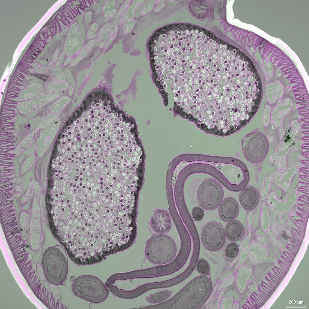
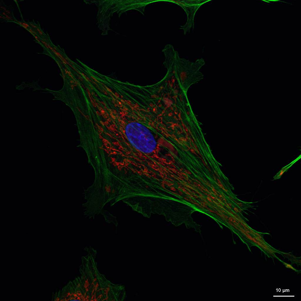
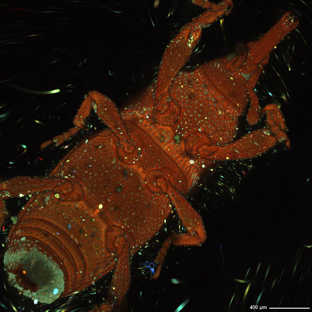
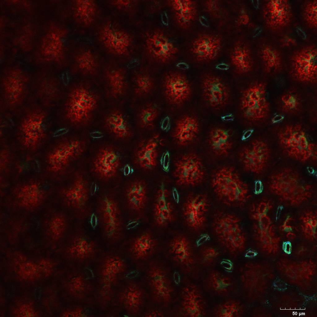
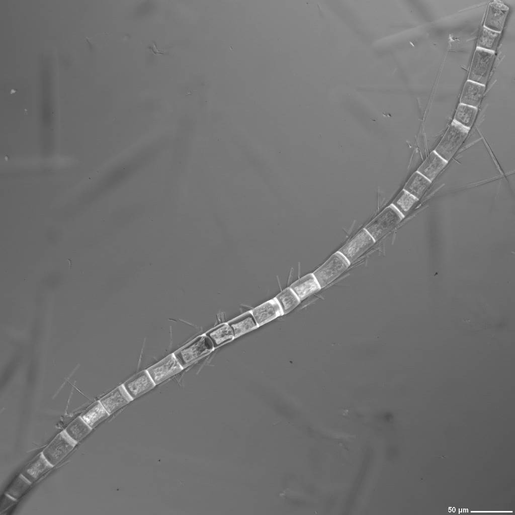
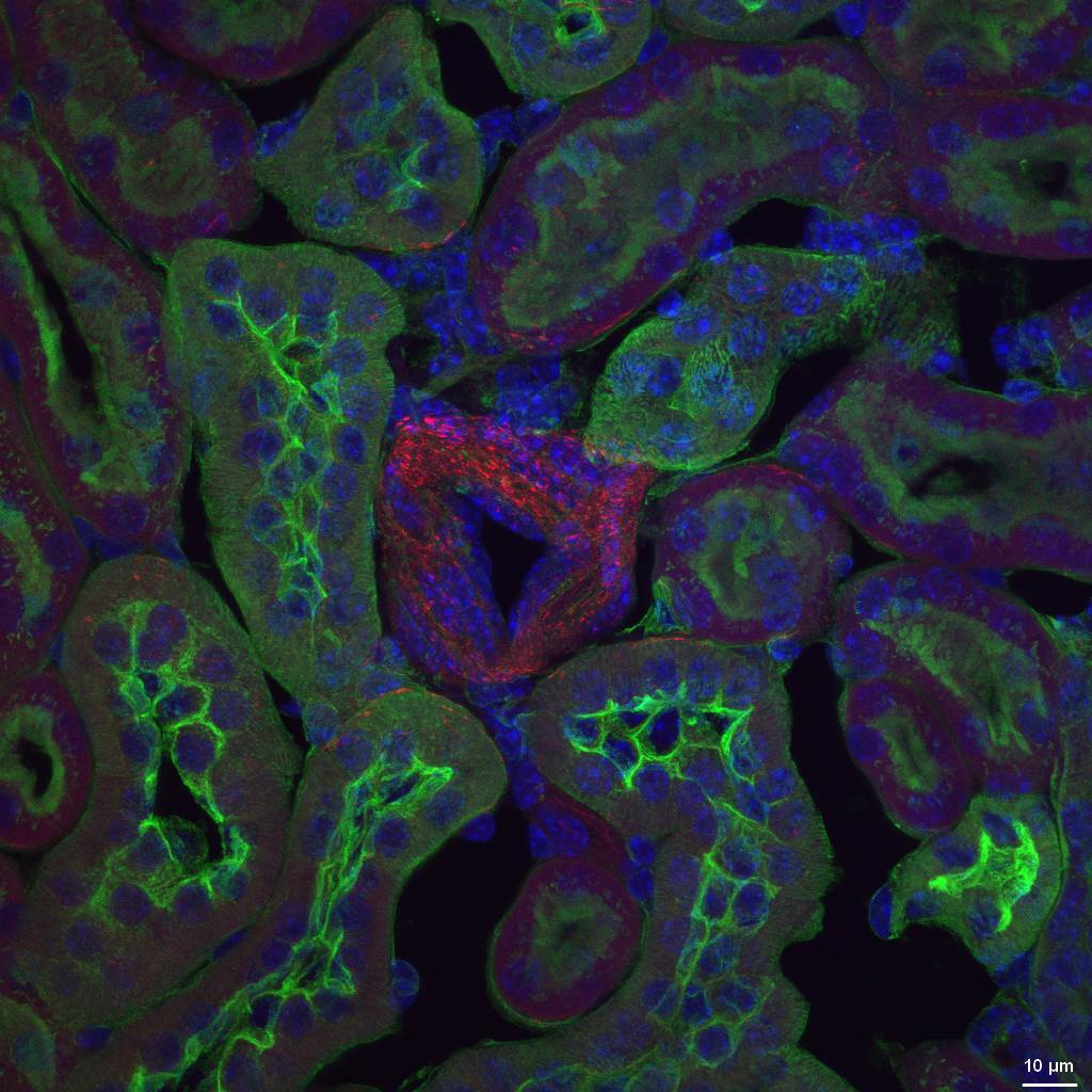
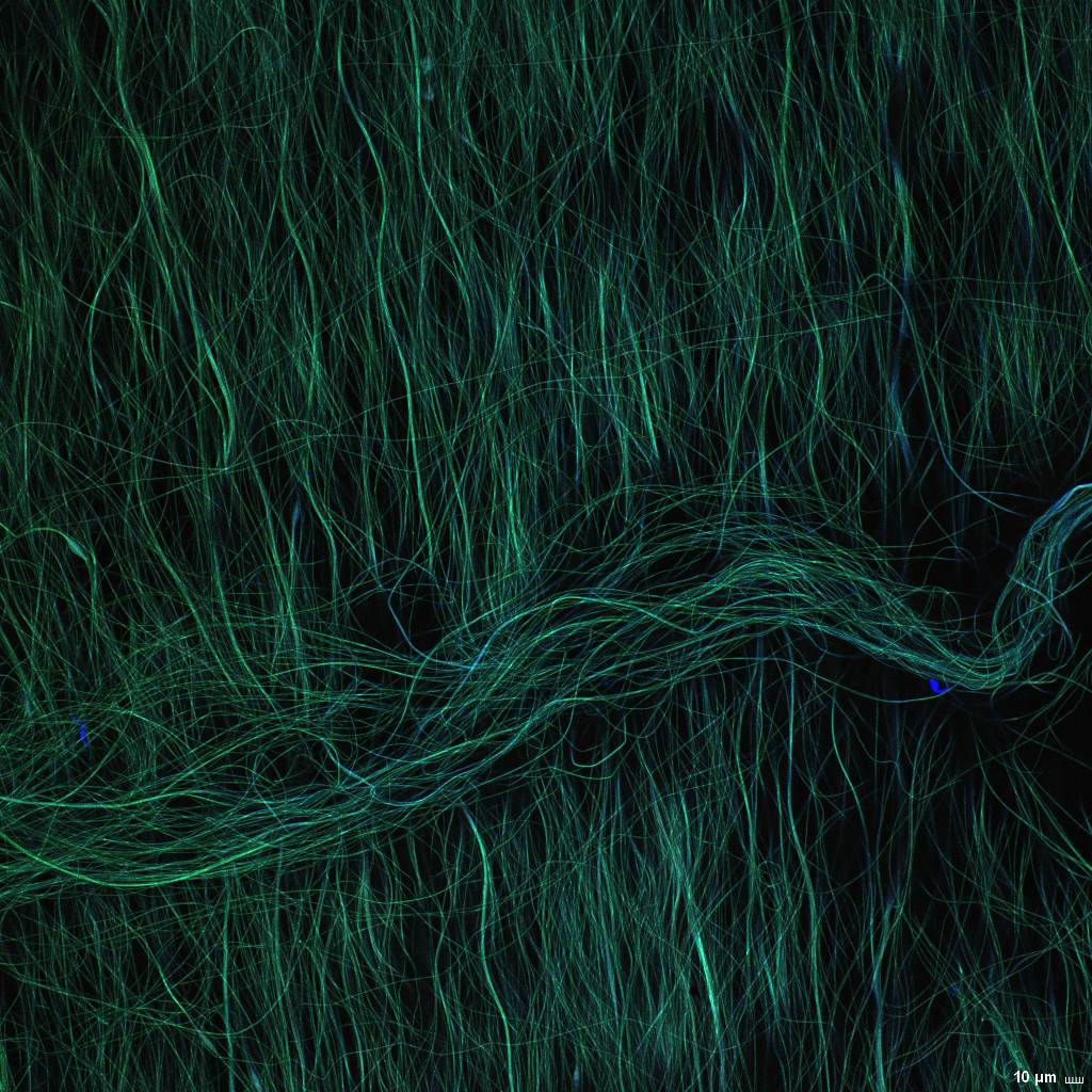
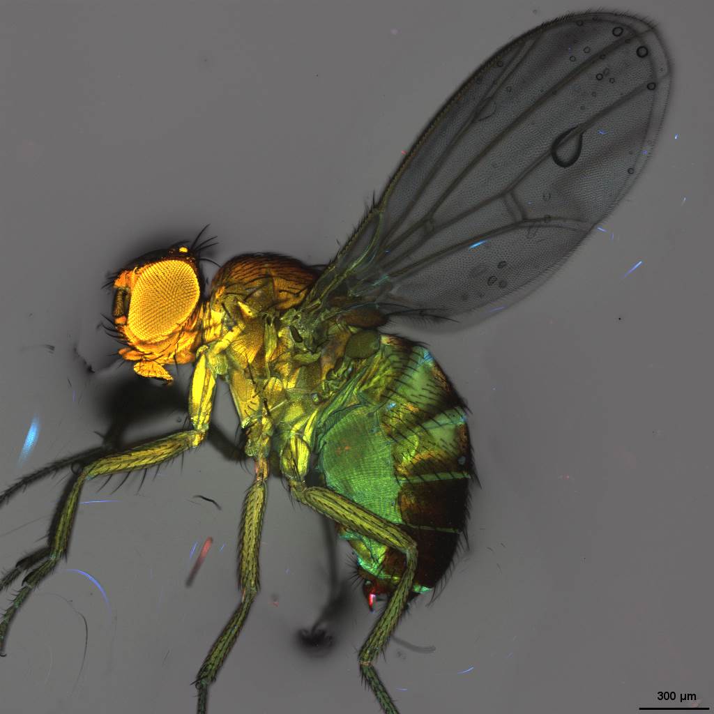
Images